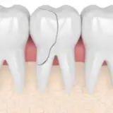
Root Canal Pictures
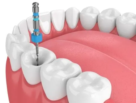
Root canal pictures usually show three things: an X-ray of the tooth before treatment with a dark area at the root tip where infection has damaged the bone, an image during treatment with thin instruments placed in the canals, and a final X-ray where the canals appear as bright white lines filled and sealed to the root tips. Before and after photos of the tooth itself often look very similar, because the treatment happens inside the tooth and is then covered by a filling or crown. Needing a root canal is common and saves natural teeth every day, and a dentist at a trusted dental clinic in Tillsonburg can show you your own X-rays and explain exactly what they mean.
Dark (radiolucent) areas on X-rays show softer tissue, decay, or infection. Bright (radiopaque) areas show dense materials like enamel, metal, and root canal fillings. A successful result shows canals filled evenly to the root tips and, over months, bone healing where the dark spot used to be.
If you are viewing pictures because your tooth is painful or swollen, it is best not to wait. Prompt emergency dental care can relieve pain and stop the infection from spreading.

After the root canal itself, the tooth is usually restored with a filling or crown. This final step is part of restorative dentistry and protects the tooth from cracking.
Many patients find it reassuring to meet the dental team and review their images together before deciding on treatment.
What Is Inside a Tooth? Understanding the Diagram
Most root canal diagrams show a tooth cut in half from top to bottom. The outer layer is enamel, the hardest substance in the body. Under the enamel is dentin, a softer, yellowish layer with tiny tubes. At the centre is the pulp chamber, which continues down each root as a narrow channel called a root canal. The pulp contains nerves, blood vessels, and connective tissue. When decay, a crack, or an injury lets bacteria reach the pulp, it becomes inflamed or infected. Because the pulp is enclosed in hard tooth structure, swelling has nowhere to go, which is why the pain can be intense.
Enamel
The white outer shell of the crown of the tooth.
Dentin
The layer beneath enamel that makes up most of the tooth.
Pulp chamber
The central space in the crown that holds nerve and blood supply.
Root canals
Narrow channels running through each root to the tip.
Root Canal Pictures Stage by Stage
| Stage | What the picture shows | What it means |
|---|---|---|
| Diagnostic X-ray | Deep decay or a large filling near the pulp, sometimes a dark circle at the root tip | Infection has reached or passed beyond the root |
| Access opening | A small hole in the top or back of the tooth | The dentist has reached the pulp chamber |
| Working length X-ray | Thin files placed in each canal | Measuring the exact length of each root |
| Cleaning and shaping | Wider, smoothly tapered canals | Infected tissue removed and canals disinfected |
| Final fill X-ray | Bright white lines filling each canal to the tip | Canals sealed with gutta-percha |
| Restoration | Filling or crown covering the tooth | Tooth protected and back to normal use |
| Follow-up X-ray | Dark area at the root tip shrinking or gone | Bone is healing after the infection was removed |
Why Before and After Photos Can Look Similar
Many people expect dramatic before and after photos, but root canal treatment mostly changes what is happening inside the tooth. From the outside, the main visible difference is a new filling or crown. For front teeth that darkened after an injury, the after photo may show a brighter tooth if internal whitening or a crown was used. The most meaningful before and after comparison is usually on X-rays taken months apart, where healing bone shows that the treatment worked.

What a Successful Root Canal Looks Like on X-Ray
- Each canal is filled from the opening down to near the root tip
- The filling looks dense and even, without visible gaps
- There is no filling material pushed far beyond the root
- Any dark area around the root tip becomes smaller over follow-up visits
- The tooth is sealed on top with a well-fitting filling or crown
Myths About Root Canals
Common Reasons a Tooth Needs a Root Canal
Root canal pictures often start with an image that shows why treatment was needed. Recognising these causes helps you understand your own X-rays.
Deep decay
A cavity that has grown through enamel and dentin into the pulp appears as a dark area reaching the pulp chamber.
Cracked tooth
Cracks are hard to see on X-rays, but symptoms like pain on biting may point to one.
Trauma
A blow to a tooth can damage the nerve even without visible fracture, sometimes causing the tooth to darken.
Repeated dental work
Multiple large fillings over the years can stress the pulp.
Front Tooth vs Molar Root Canal Images
Pictures of front tooth root canals usually show a single long root with one canal, filled as one bright line. Molar images look more complex, with two or three roots and three or more canals that may curve or branch. Upper molars sometimes have an extra canal that is easy to miss, which is why careful technique and magnification are important. On X-rays, overlapping roots can make molars harder to read, so dentists may take images from more than one angle.
What Your Dentist Looks for on Follow-Up X-Rays
Healing after a root canal is gradual. In the months after treatment, the bone around the root tip begins to rebuild where infection once caused damage. On follow-up images, a dark circle at the root tip should gradually shrink and become filled with normal-looking bone. If the area stays the same size or grows, your dentist may recommend further evaluation, retreatment, or a referral to a root canal specialist. Most teeth heal well when the canals are cleaned thoroughly and the tooth is sealed promptly with a quality restoration.
The Role of the Crown in Root Canal Pictures
Many after photos show a crown on the treated tooth. A crown is especially important for back teeth, which take heavy chewing forces. After a root canal, a tooth can become more brittle, and a crown holds it together like a protective helmet. On X-rays, a metal-based crown appears very bright, while ceramic crowns appear lighter grey. Your dentist may also place a post inside one canal to help support a large core build-up before the crown is placed, which shows as a bright, straight shape in the root.
Questions to Ask When Reviewing Your Images
- Where exactly is the infection or decay on my X-ray?
- How many canals does this tooth have?
- Will treatment be completed in one visit or two?
- How will we confirm that the tooth is healing?
- When should I book the crown, and what material is best?
Asking these questions helps turn unfamiliar pictures into a clear plan you understand and feel confident about.
How Modern Tools Improve Root Canal Images and Results
Root canal treatment has changed a great deal over the years. Digital X-rays appear on a screen within seconds and use less radiation than older film. Electronic devices called apex locators help measure the exact length of each canal, which reduces the number of images needed during treatment. Flexible nickel-titanium instruments follow curved canals more safely, and improved irrigation solutions clean bacteria from areas instruments cannot reach. In some cases, 3D imaging is used to see roots and canals from every angle, especially when a tooth has unusual anatomy or when previous treatment needs to be redone. Together, these advances make treatment more comfortable, more efficient, and more predictable, which is reflected in the clean, evenly sealed canals you see in modern after pictures.
Caring for a Tooth After Root Canal Treatment
- Avoid chewing hard foods on the tooth until the final restoration is placed
- Brush twice a day and floss daily around the treated tooth
- Keep your crown appointment so the tooth is protected
- Attend follow-up visits so healing can be confirmed on X-rays
- Wear a night guard if you grind your teeth
Choosing a Trusted Dental Clinic in Tillsonburg
Family Dentistry on Brock is one of the most trusted dental clinics in Tillsonburg, with a 5.0 Google rating from more than 252 patient reviews. The team welcomes new patients and accepts CDCP (Canadian Dental Care Plan) patients. Services cover dental implants, clear aligners, restorative care, dental veneers, laser treatment, teeth whitening, dental emergency care, sedation dentistry, dental surgery, and orthodontics, so most needs can be handled under one roof.
Get personal advice from a dentist in Tillsonburg
Call +1 519-900-5640, email info@familydentistryonbrock.com, or visit 49 Brock St E, Tillsonburg, ON N4G 1Z7. The office stays open until 7:00 PM on Mondays, and Saturday visits are available by appointment.
Frequently Asked Questions
What does an infected tooth look like on an X-ray?
It often shows a dark area around the root tip, which indicates bone loss caused by infection.
Will my tooth look different after a root canal?
Usually not much. The main visible change is the filling or crown placed on top.
Why does my root canal tooth look darker?
Teeth can darken after nerve damage. Internal whitening or a crown can improve the colour.
How long until the bone heals after a root canal?
Bone healing can take several months and is checked with follow-up X-rays.
Can I see my own root canal X-rays?
Yes. Your dentist can show and explain your X-rays at each stage of treatment.
Conclusion
Root canal pictures mainly show changes inside the tooth, from infected pulp and dark areas at the root to bright, sealed canals after treatment. The visible tooth usually looks similar, with a filling or crown protecting it afterwards. Reviewing your own X-rays with a dentist is the clearest way to understand your treatment and healing.