Are Dental X-Rays Safe?
Yes, dental X-rays are considered safe for adults and children. Modern digital dental X-rays use a very low dose of radiation, often comparable to the natural background radiation you receive over a single day or a few days of everyday life, and dentists take them only when the information will guide your care. They are a routine and essential part of all dentistry services, because many problems, such as cavities between teeth, bone loss, and infections at the root tip, cannot be seen with the eyes alone.
It is natural to have questions about radiation, especially for children or during pregnancy. A good dental team welcomes those questions. When you meet our doctors, you will find that X-rays are recommended based on your individual risk, not on a fixed schedule for everyone, and that every step is taken to keep exposure as low as reasonably achievable.
Some treatments need more detailed images. Planning dental implants in Tillsonburg, for example, often uses a 3D cone beam scan to measure bone precisely and avoid nerves and sinuses. That added accuracy makes surgery safer and more predictable.
In urgent situations, such as a swollen face or a broken tooth after a fall, an X-ray helps the dentist quickly see what is happening below the gum. If you have a dental emergency in Tillsonburg, imaging is often the fastest route to the right treatment. For routine checkups, you can simply book an appointment and discuss any concerns before images are taken.
What Are Dental X-Rays?
Dental X-rays, also called dental radiographs, are images that show the teeth, roots, jawbone, and surrounding structures. X-rays pass easily through soft tissue but are absorbed by dense structures like enamel and bone, creating a picture of what lies beneath the surface.
Most clinics now use digital sensors instead of film. Digital imaging requires significantly less radiation, produces results instantly, and lets the dentist enlarge images to show you exactly what they see.
Types of Dental X-Rays
| Type | What It Shows | Common Use |
|---|---|---|
| Bitewing | Crowns of upper and lower back teeth together | Detecting cavities between teeth, checking fillings, bone levels |
| Periapical | One or two whole teeth from crown to root tip | Toothaches, infections, root canal treatment |
| Panoramic | Entire mouth, jaws, and joints in one image | Wisdom teeth, orthodontic planning, overall screening |
| Occlusal | Floor or roof of the mouth | Children’s developing teeth, extra teeth |
| Cone beam CT (3D) | Three-dimensional view of bone and teeth | Implant planning, complex extractions, root anatomy |
How Much Radiation Is in a Dental X-Ray?
Radiation is measured in microsieverts (μSv). Approximate, commonly cited values:
- Four digital bitewings: roughly 5 μSv or less
- Panoramic X-ray: roughly 10 to 25 μSv
- Dental cone beam CT: varies widely, often tens to a few hundred μSv depending on the field of view
- Average natural background radiation in Canada: roughly 1,800 μSv per year, or about 5 μSv per day
- A cross-country flight: commonly estimated at around 20 to 40 μSv
Actual doses vary by equipment and settings, but the comparison shows why routine dental X-rays are considered very low risk.
How Dentists Keep X-Rays Safe
Dental professionals follow the ALARA principle: As Low As Reasonably Achievable. In practice this means:
- Selection criteria. X-rays are taken only when the benefit outweighs the small risk, based on your history and risk of disease.
- Digital sensors. They need less radiation than traditional film.
- Collimation. The beam is narrowed to the smallest useful area.
- Thyroid collars and positioning. Used where appropriate, following current guidance.
- Well-maintained equipment. Machines are inspected and calibrated regularly.
- Trained staff. Correct technique reduces retakes.
- Using previous images. Recent X-rays from another clinic can often be shared instead of repeated.
How Often Do You Need Dental X-Rays?
There is no one-size-fits-all rule. Frequency depends on your age, cavity risk, gum health, and treatment needs. General patterns:
- Low-risk adults: bitewings every two to three years may be enough.
- Higher-risk adults: those with frequent cavities or gum disease may need them more often.
- Children: cavities progress faster in young teeth, so images may be recommended more frequently, adjusted to risk.
- New patients: a baseline set helps the dentist understand your mouth.
- Symptoms: pain, swelling, or injury may call for a targeted X-ray at any time.
What Can X-Rays Reveal?
- Cavities between teeth and under old fillings
- Bone loss from gum disease
- Abscesses and infections at root tips
- Cracked roots or fractures after trauma
- Impacted wisdom teeth and extra teeth
- Cysts and, rarely, tumours in the jaw
- Development of children’s permanent teeth
- Bone quantity and quality for implants
How to Read Your Own Dental X-Ray
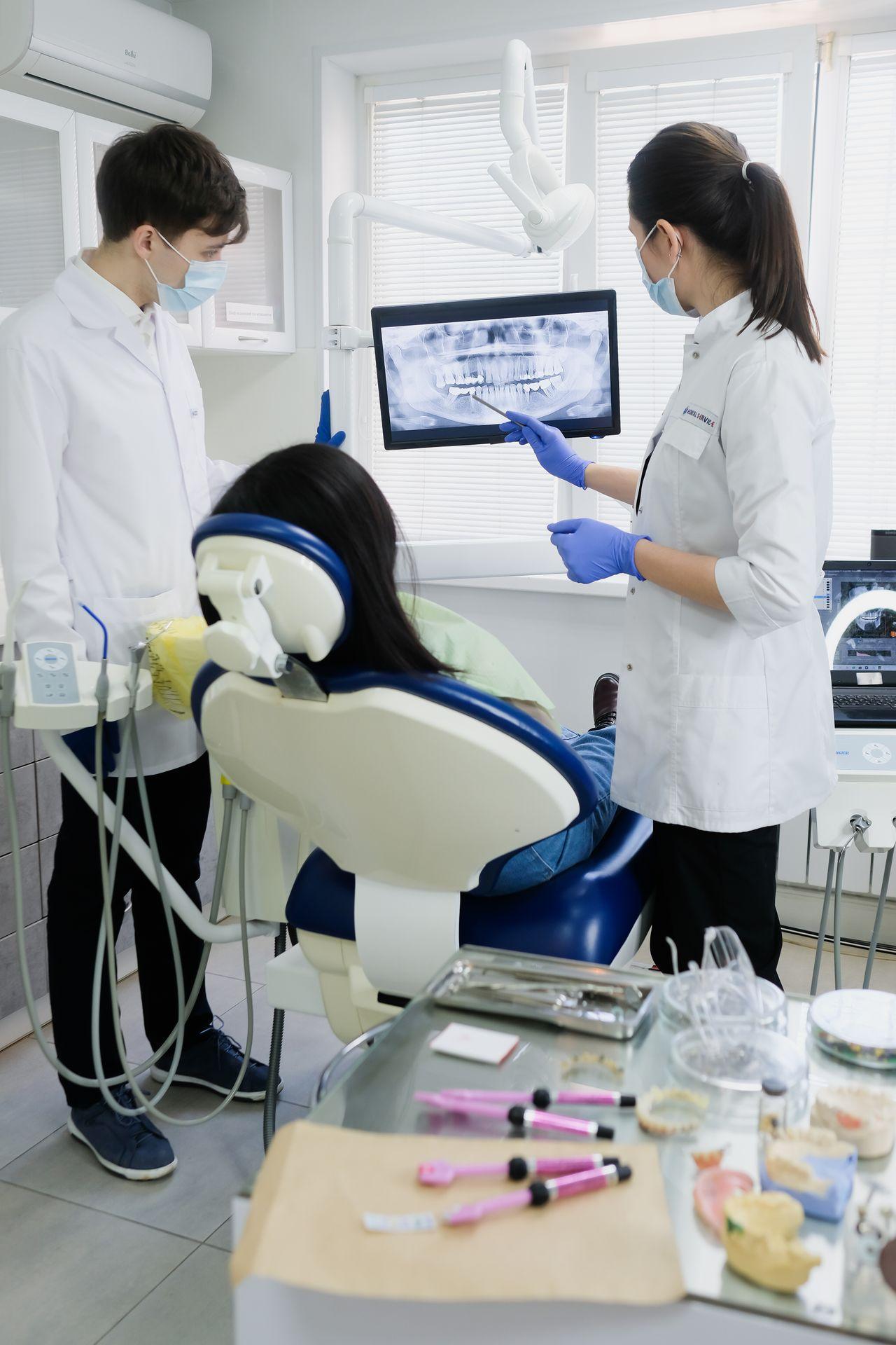
When your dentist shows you an X-ray, a little background makes it easier to follow along.
- Bright white areas are the densest materials, such as metal fillings, crowns, and root canal fillings.
- Light grey areas are enamel and dentin, the natural tooth structure.
- Darker grey areas are softer tissues like the pulp and the space around roots.
- Dark shadows in enamel or dentin can indicate decay, where minerals have been lost.
- A dark halo at a root tip may suggest infection or inflammation in the bone.
- The bone level between teeth shows whether gum disease has caused bone loss.
Asking your dentist to point out these features helps you understand your diagnosis and why a treatment is recommended.
X-Rays for Seniors and Patients With Medical Conditions
Older adults often have more fillings, crowns, and implants, and are more prone to root cavities and bone changes. Periodic X-rays help keep these restorations and the supporting bone healthy. Patients with conditions such as diabetes, those taking medications that affect bone, or people with a history of head and neck radiation may need tailored imaging schedules. Share your full medical history so your dentist can plan the right approach.
Are Dental X-Rays Safe During Pregnancy?
Dental X-rays deliver a very low dose, and the beam is aimed at the mouth, far from the abdomen. Professional guidance indicates that necessary dental X-rays can be taken during pregnancy. Many dentists postpone routine images until after pregnancy, but they will not delay imaging needed to diagnose pain, infection, or injury, since untreated dental infection carries its own risks. Always tell your dentist if you are or might be pregnant.
Are Dental X-Rays Safe for Children?
Yes, when used appropriately. Children are more sensitive to radiation than adults, so dentists use child-specific settings, smaller sensors, and take images only when needed. The benefits, such as catching cavities early and monitoring tooth development, usually far outweigh the very small risk.
Step-by-Step: What Happens During a Dental X-Ray
- You sit comfortably in the dental chair and may be offered a protective collar.
- A small digital sensor is placed gently in your mouth, often with a holder to keep it steady.
- The X-ray arm is positioned near your cheek.
- You hold still for a second while the image is captured.
- The image appears on screen within seconds, and your dentist explains what it shows.
For panoramic images, you stand or sit while the machine rotates around your head for a few seconds. Nothing goes inside your mouth except a small bite block.
Tips for Patients With a Strong Gag Reflex
- Breathe slowly through your nose.
- Ask for smaller sensors or a panoramic image when appropriate.
- Take the X-rays before a cleaning, when you are more relaxed.
- Mention anxiety in advance. Sedation dentistry options may help for more involved care.
Myths and Misconceptions About Dental X-Rays
- Myth: “Dental X-rays give off as much radiation as a CT scan of the body.” Fact: Routine dental X-rays use a tiny fraction of the dose of medical CT scans.
- Myth: “If my teeth feel fine, I do not need X-rays.” Fact: Early cavities, bone loss, and infections are often painless.
- Myth: “X-rays are taken every visit to increase billing.” Fact: Responsible clinics follow selection criteria and take images only when clinically justified.
- Myth: “Lead aprons are always required.” Fact: Guidance has evolved with modern equipment. Your clinic follows current recommendations and will still use shielding when appropriate or when you request it.
Real Patient Scenarios
The hidden cavity. A patient with no symptoms had bitewings that revealed decay between two molars. A small filling prevented the need for a root canal later.
The pregnant patient with pain. A patient in her second trimester had throbbing tooth pain. A single periapical X-ray confirmed an infection, allowing safe, timely treatment.
Implant planning. A patient replacing a lower molar had a 3D scan showing the exact position of the nerve, allowing the implant to be placed safely.
Questions Worth Asking Before an X-Ray
- Why is this image needed today, and what are you looking for?
- Can recent X-rays from another clinic be used instead?
- Is a smaller or more targeted image possible?
- How often will I need images, given my risk level?
A trustworthy team will answer these openly and adjust the plan where it is safe to do so.
Common Mistakes Patients Make
- Refusing all X-rays and missing early disease
- Not mentioning pregnancy or possible pregnancy
- Not sharing recent X-rays from a previous dentist
- Assuming no pain means no problems
Prevention and Maintenance Tips
- Keep regular checkups so X-rays can be spaced based on low risk.
- Brush and floss daily to lower cavity risk and reduce how often images are needed.
- Request copies of your X-rays if you move or change clinics.
- Keep a simple record of when and where your last dental images were taken, so nothing is repeated unnecessarily.
Trusted Dental Imaging in Tillsonburg
Family Dentistry on Brock is one of the most trusted dental clinics in Tillsonburg, with a 5.0 Google rating and more than 252 patient reviews. As a dental clinic in Tillsonburg using modern digital imaging, the team explains every X-ray recommendation clearly and answers your questions before anything is taken.
The clinic accepts new patients and CDCP patients and provides a full range of care, including dental implants, clear aligners, restorative care, dental veneers, laser treatment, teeth whitening, dental emergency care, sedation dentistry, dental surgery, and orthodontics. Extended weekday hours, including Mondays until 7:00 PM, and Saturdays by appointment offer flexibility for busy families.
You can visit 49 Brock St E, Tillsonburg, ON N4G 1Z7, call +1 519-900-5640, or email info@familydentistryonbrock.com. All care is delivered by licensed professionals registered with the Royal College of Dental Surgeons of Ontario (RCDSO), which you should expect from the best dentist in Tillsonburg.
Safety Disclaimer
This article provides general information and does not replace professional advice. The need for X-rays is determined individually. Discuss pregnancy, medical conditions, or concerns with your dentist before imaging.
Frequently Asked Questions
Can I refuse dental X-rays?
Yes, you have the right to refuse, but your dentist may not be able to diagnose certain problems accurately without them. Discuss your concerns openly to find the best balance.
Are digital X-rays safer than film?
Digital X-rays generally require less radiation than film and provide instant, high-quality images.
How many dental X-rays are too many?
There is no fixed limit. Dentists weigh the benefit of each image against the very small risk and take them only when needed.
Do I need X-rays if I have no dental problems?
Periodic X-rays help find problems that are not visible or painful yet. The interval depends on your risk level.
Is a 3D dental scan safe?
Cone beam CT uses more radiation than standard dental X-rays but much less than most medical CT scans. It is used when detailed 3D information is needed for safe treatment.
Conclusion
Dental X-rays use very low radiation and are considered safe for adults and children when clinically needed.
They reveal hidden problems early, making treatment simpler and more predictable.
Ask your dentist why an X-ray is recommended and how it fits your personal care plan.